Interested in using this facility?
For all enquiries please contact:
General Manager of the Imperial College Clinical Imaging Facility
Dr Albert Busza
a.busza@imperial.ac.uk
+44 (0)20 7594 0996
Terms and conditions are available on request.
The Imperial College Clinical Imaging Facility (CIF) provides support for interdisciplinary research activities across the university, integrating the physical, biological and medical sciences.
We provide the capability to investigate a wide range of clinical conditions using magnetic resonance imaging (MRI), Positron Emission Tomography (PET), electroencephalography (EEG) and transcranial brain stimulation. We have expertise in neuroscience and oncology, as well as multimodal imaging. We are part of the Faculty of Medicine at Imperial and are located at the Hammersmith Hospital, benefiting from the hospital’s long tradition of world-class imaging research.
We currently support a broad range of applied research studies and have strong links to academics and clinicians as well as commercial organisations. We welcome research proposals from all academic researchers and industry.
“The last decade has seen amazing advances in imaging technology. We can now use imaging to accurately map connections in the brain, track the development of dementia and guide cancer treatment. In the Clinical Imaging Facility we aim to translate these technological advances into improved diagnosis and treatments.
Professor David Sharp, Scientific Director of the Imperial College Clinical Imaging Facility
Discover our facility
Watch our video to hear directly from our researchers about the significance of the clinical imaging facility and its application in medical science and innovation.
Imaging resources

Magnetic Resonance Imaging (MRI)
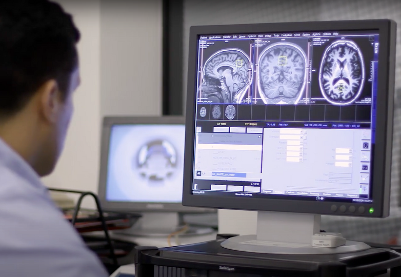
The Siemens 3T Verio MRI scanner comprises a 70cm diameter open-bore, short-axis magnet. It has been equipped with a comprehensive range of transmit/receive coils suitable for many body regions and applications. The relatively large bore (compared to most MRI systems) ensures a higher degree of subject comfort and compliance.
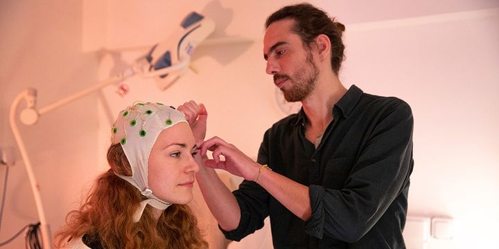
Electroencephalography (EEG)
32-channel EEG system compatible with functional MRI. We have a BrainProducts 32-channel amplifier (BrainAmp MRplus) an additional 8-channel bipolar amplifier (BrainAmp ExG MR), which can be used to record electrooculogram, motor evoked potentials, etc. The system is equipped with a SyncBox to synchronize the EEG and MRI scanner clocks and a trigger cable to send TTL triggers to the EEG system from a computer running a behavioural task.
Positron Emission Tomography (PET)
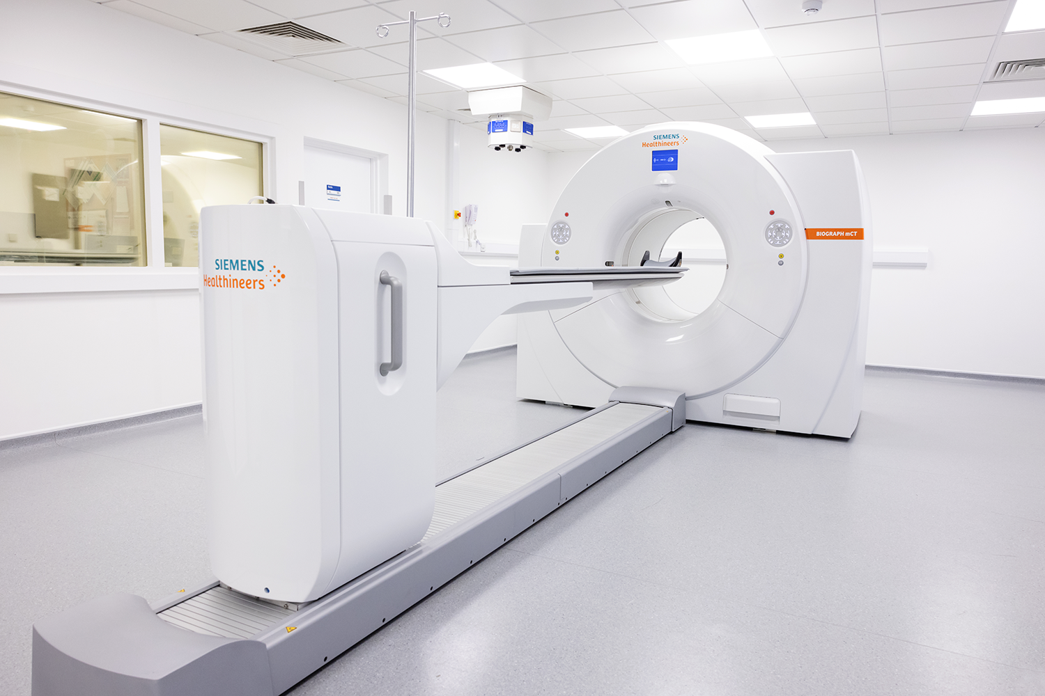
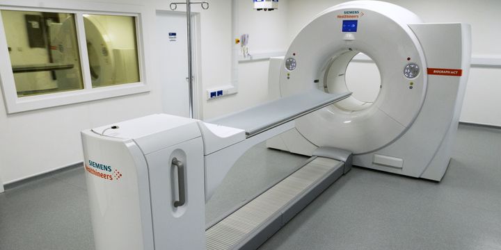
The Siemens Biograph mCT-S 64 is a sophisticated whole-body PET-CT scanner designed for oncological, neurological, and cardiac imaging. It offers a non-invasive method to produce high-quality CT and PET images, revealing detailed anatomical and molecular biological processes. Key features include:
- High-performance 64 slice spiral CT imaging.
- High-resolution PET imaging for metabolic and physiological assessments with 3D iterative reconstruction.
- Effective registration of anatomical and metabolic images for precise lesion detection.
- Advanced attenuation and scatter correction for improved PET image quality with UltraHD PET.
The Biograph mCT-S 64 features a fully integrated PET-CT gantry that combines CT and PET detectors in a compact design, minimising data transmission noise and enhancing reliability. Its large gantry opening, and short tunnel length accommodate patients weighing up to 227 kg and reduce feelings of claustrophobia. The system also includes quad operator controls for easy positioning and dual displays for monitoring system status from both sides of the patient.
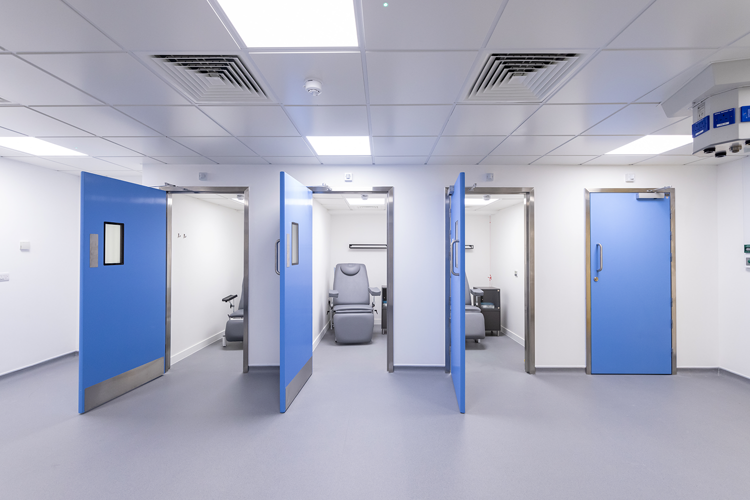
An Allogg Automated Blood Sampling System (ABSS) is available for those studies which require arterial blood sampling; there is also an adjacent tissue/blood processing lab with dual-HPLC/radioactivity detectors for tracer metabolite analysis.
Transcranial Electric Stimulation (TES)
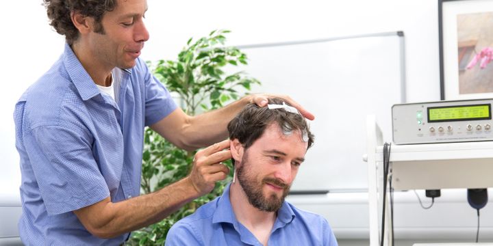
MR-compatible stimulators (DC stimulator MR) from neuroConn. These are used to deliver non-invasive brain stimulation in humans, using the following TES techniques:
- tDCS – transcranial direct current stimulation
- tACS – transcranial alternating current stimulation
- tRNS – transcranial random noise stimulation
Our devices can be controlled remotely using data acquisition (DAQ) devices.
Gallery
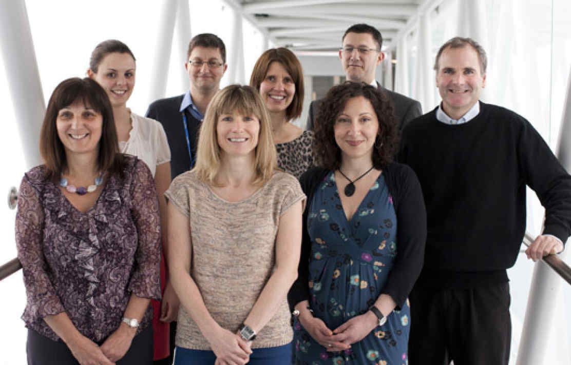
Meet the team
 |
Professor David Sharp |
 |
Dr Albert Busza |
 |
Pedro Vicente |
 |
Rob Punjani |
