X-ray Crystallography: A Very Brief History
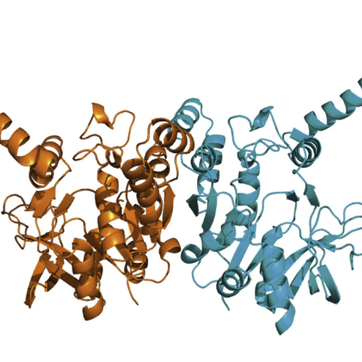
Macromolecular X-ray crystallography is a structural biology technique used to determine the atomic structure of macromolecules such as proteins, DNA, and RNA. The origins of X-ray crystallography begin in 1912, when Max von Laue exposed a copper sulphate crystal to X-rays to produce a diffraction pattern. This proved the theory X-rays were electromagnetic waves, that the regular spacing of the atoms in a crystal could act as a diffraction grating, and the atomic theory of matter earning von Laue the Nobel prize in Physics in 1914.
Building on this work the father son duo William and Lawrence Bragg were responsible for the birth of X-ray crystallography in 1913. Using an X-ray spectrometer build by William Bragg and Bragg's law of X-ray diffraction determined by Lawrence Bragg, the pair solved the crystal structures of sodium chloride, potassium chloride, potassium bromide and potassium iodide in their paper "The structure of some crystals as indicated by their diffraction of X-rays". in 1915 they were both awarded the Nobel prize in Physics, making Lawrence the youngest person ever to receive the accolade.
It would be many years until the first protein structure was solved, a feat achieved by john Kendrew in 1958 with the structure of myoglobin. A structure 25 years in the making which heralded the beginning of Protein Crystallography. Fast-forward to the modern day and macromolecular crystallography has become an essential tool for biologists to determine the structure of significantly more complex proteins and protein complexes. The considerable leaps forward in computing power, synchrotrons, detectors, molecular biology, and robotics means structure solution is easier today than it has ever been, but still not a given. If you have pure protein you too could become a crystallographer.
The prolific work of structural biologists for the past 60+ years has resulted in over 200,000 structures being deposited into the Protein Data Bank (PDB), a huge dataset which has fueled the development of protein prediction software like Aplhafold which is now in it's thrid generation.
Crystallisation
Condiserable advances in the molecular biology of recombinant protein expression and the addition of affinity tags to proteins of interest means that crystallisation has now become the major bottle neck in the protein crystallography pipeline. Protein crystallisation is achieved by addition of mild precipitation agents to purified protein solution in the right concentration to produce a supersaturated state which encourages the macromolecules to fall out of solution and form a crystal.
The most common method for macromolecular crystallisation is vapour diffusion. In this method the protein and precipitant are mixed in a sealed enviroment above an excess of the precipitation solution, also known as mother liquor or well solution. Over time the protein/precipitate drop and well solution equillibrate, with water diffusing from the mixed drop to the well solution. This causes an increase in the protein and precipitant concentration, which can lead to nucleation of protein crystals. As crystals begin to form the protein concetration in solution decreases, and nucleation of new crystals will stop allowing for growth of the existing crystals. These crystallisation trajectories are best explained through use of a phase diagram, such as those produced in Beale et al 2019 and Stohrer et al 2021.
Finding the right condition to form crystals can be challenging and sample intensive. However, the development of robotics and commercial screens which cover a broad range of chemical space has simplified the process of finding and optimising these conditions for your protein while keeping sample consumption to a minimum.
For a more thorough introduction to protein crystallisation I would reccomend the tutorials put together by Therese Bergfors from Uppsala University in Sweden and this Introduction to Protein Crystallisation from McPherson and Gavira 2012.
Preparing Crystals for Diffraction Experiments
Modern X-ray Sources produce high intensity X-ray beams which cause significant rediation damage to biological molecules during data collection. To mitigate this damage the majority of protein crystal diffraction data are collected at cryogenic temperatures (100 K) by flash cooling the crystals in liquid nitrogen. This increases the lifetime of the crystals in the X-ray beam to allow complete datasets, or even multiple datasets, to be collected from a single protein crystal. The major downside is the need to cryoprotect the protein crystals to prevent formation of ice crystals which can degrade diffraction. A number of common laboratory chemicals can act as cryoprotectants (glycerol, PEG 400, ethylene glycol, MPD, sucrose), but experimentation may be required to find the right one for your crystallisation condition.
Room Temperature Data Collection
Recent leaps forward in the speed of detectors and development of new data collection techniques such as the in-situ beamline VMXi, and serial crystallography means it is also possible to collect room temperature protein diffraction. This is particularly useful if you are interested in studying the dynamic properties of your protein which would be rendered immobile under cryogenic data collection.
Data processing
Processing X-ray diffraction data has never been more accessible, with the development of autoprocessing pipelines at synchrotrons which will perform data reduction and even phase your structure for you it is possible to produce a structural model with very little prior knowledge. Should the automatic pipelines fail the facility manager is available to provide guidance and training on manually processing your diffraction data and structure solution/refinement.

