Brillouin imaging is a powerful technique for non-contact and non-invasive optical imaging. This method is based on the phenomenon of Brillouin light scattering, first described by French physicist Leon Brillouin in 1922, in which light interacts with acoustic vibrations in the medium. As the result of such interaction, the light beam undergoes scattering event, in which the frequency of light beam changes by a small amount. Brillouin imaging is based on mapping these small frequency changes, known as Brollouin frequency shift, for the entire 3D object. The shift is related to microscale mechanical properties and elasticity of the object. The resolution achievable in Brillouin imaging is on the order of submicron, allowing imaging of small biological objects such as cells, membranes and tissue components. In our group we apply the principles of Brillouin microscopy to learn about disease-related modifications in elastic properties of tissues. A few examples are given below.
1. Brillouin Imaging of Arterial Wall
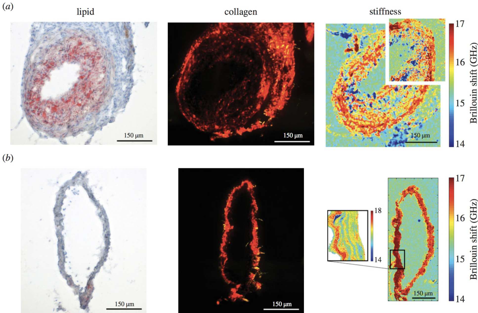
It is known that changes in the arterial wall elasticity can be related to atherosclerosis and arterial plaque formation. In the recent experimental work coming from our group, Brillouin microscopy was tested on a healthy and instrumented artery of a mouse (see Figure). The cuff positioned inside the instrumented artery induced formation of a vulnerable plaque within a few weeks. The results of Brillouin microscopy on healthy (control) and instrumented artery have shown a clear evidence of the arterial disease, fibroatheroma, and validated the measurement benefits for the disease monitoring.
Figure: images of (left) lipid (imaged with light microscopy), (centre) collagen (imaged with polarized light microscopy) and (right) Brillouin shift (imaged with Brillouin microscopy), which is related to stiffness of the (a) instrumented and (b) control vessels. Scale bars, 150 µm. Images are taken from the publication Antonacci et. al. "Quantification of plaque stiffness by Brillouin microscopy in experimental thin cap fibroatheroma" J. R. Soc. Interface 12, 20150843, 2015.
2. In-vitro Brillouin Imaging of Single Cells
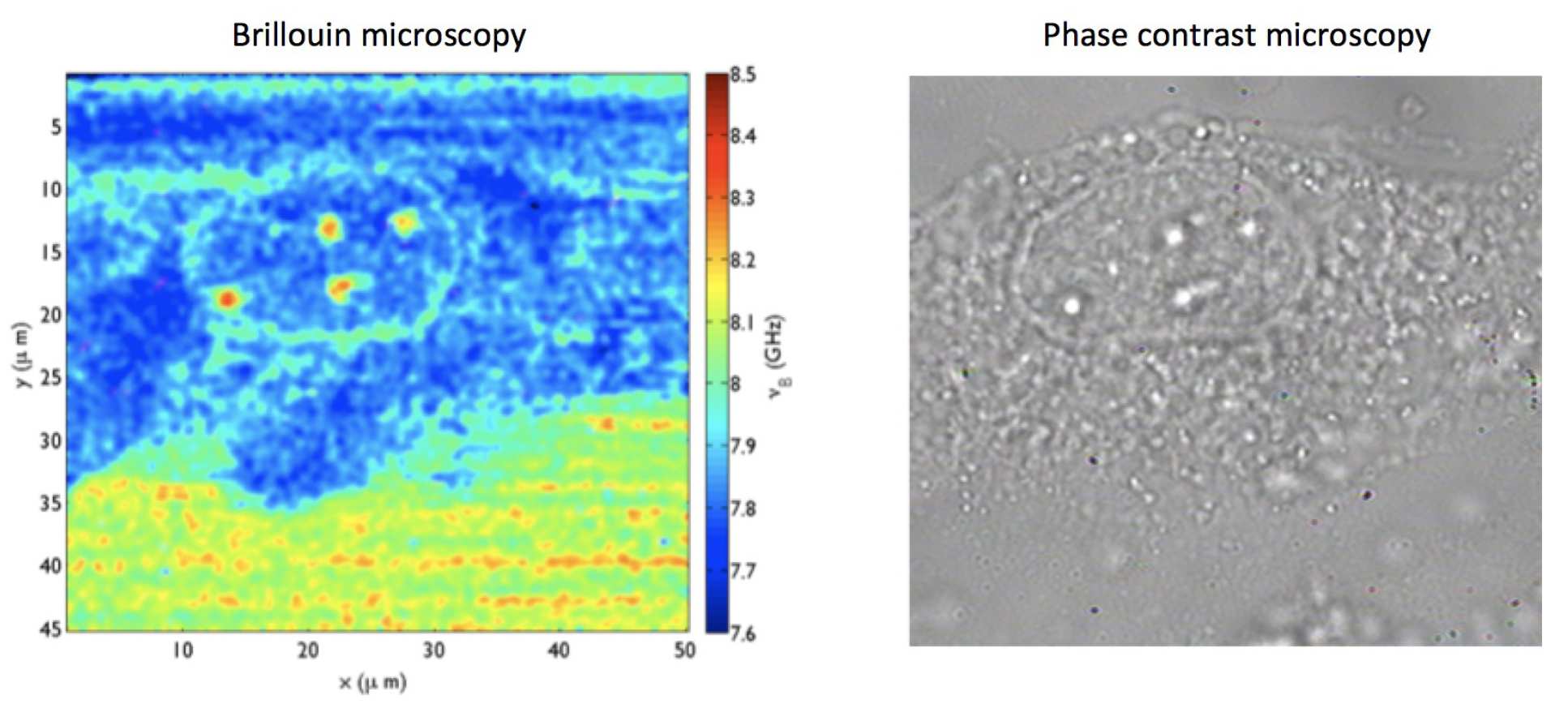
Stiffness variation of endothelial cells might be responsible for Glaucoma. In-vitro Brillouin imaging in capable of detecting individual nucleoli inside the nucleus of a single cell (see figure below) and shows that nucleoli are stiffer than the nucleus.
Figure: endothelial cells from Schlemm's canal. These images present the word’s first sub-cellular resolution Brillouin images. This work is done in collaboration with Dr. Overby and S. Braakman from Imperial College Bioengineering Department. Images are credit to Guiseppe Antonacci.
