Main content blocks

About us
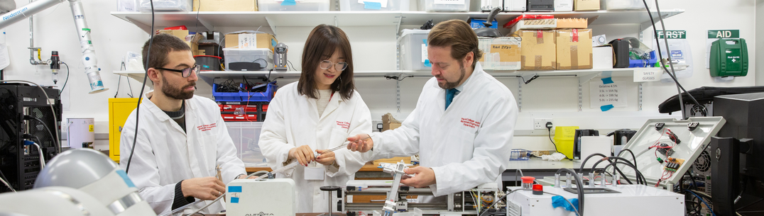
The MIM Lab develops robotic and mechatronics surgical systems for a variety of procedures.
-
Twitter/X: @MIMLab_Robotics
-
Youtube: Mechatronics in Medicine Lab
Research lab info
What we do
The Mechatronics in Medicine Laboratory develops robotic and mechatronics surgical systems for a variety of procedures including neuro, cardiovascular, orthopaedic surgeries, and colonoscopies. Examples include bio-inspired catheters that can navigate along complex paths within the brain (such as EDEN2020), soft robots to explore endoluminal anatomies (such as the colon), and virtual reality solutions to support surgeons during knee replacement surgeries.
Why it is important?
...
How can it benefit patients?
......
Meet the team
Results
- Showing results for:
- Reset all filters
Search results
-
Journal articleFranco E, Rodriguez y Baena F, Astolfi A, 2020,
Robust dynamic state feedback for underactuated systems with linearly parameterized disturbances
, International Journal of Robust and Nonlinear Control, Vol: 30, Pages: 4112-4128, ISSN: 1049-8923This article investigates the control problem for underactuated port‐controlled Hamiltonian systems with multiple linearly parameterized additive disturbances including matched, unmatched, constant, and state‐dependent components. The notion of algebraic solution of the matching equations is employed to design an extension of the interconnection and damping assignment passivity‐based control methodology that does not rely on the solution of partial differential equations. The result is a dynamic state‐feedback that includes a disturbance compensation term, where the unknown parameters are estimated adaptively. A simplified implementation of the proposed approach for underactuated mechanical systems is detailed. The effectiveness of the controller is demonstrated with numerical simulations for the magnetic‐levitated‐ball system and for the ball‐on‐beam system.
-
Conference paperHu X, Fabrizio C, Tatti F, et al., 2020,
Automatic calibration of commercial optical see-through head-mounted displays for medical applications
, 2020 IEEE Conference on Virtual Reality and 3D User Interfaces, Publisher: IEEE, Pages: 754-755The simplified, manual calibration of commercial Optical See-Through Head-Mounted Displays (OST-HMDs) is neither accurate nor convenient for medical applications. An interaction-free calibration method that can be easily implemented in commercial headsets is thus desired. State-of-the-art automatic calibrations simplify the eye-screen system as a pinhole camera and tedious offline calibrations are required. Furthermore, they have never been tested on original commercial products. We present a gaze-based automatic calibration method that can be easily implemented in commercial headsets without knowing hardware details. The location of the virtual target is revised in world coordinate according to the real-time tracked eye gaze. The algorithm has been tested with the Microsoft HoloLens. Current quantitative and qualitative user studies show that the automatically calibrated display is statistically comparable with the manually calibrated display under both monocular and binocular rendering mode. Since it is cumbersome to ask users to perform manual calibrations every time the HMD is re-positioned, our method provides a comparably accurate but more convenient and practical solution to the HMD calibration.
-
Journal articleVirdyawan V, Dessi O, Rodriguez y Baena F, 2020,
A novel sensing method to detect tissue boundaries during robotic needle insertion based on laser Doppler flowmetry
, IEEE Robotics and Automation Letters, Vol: 5, Pages: 1524-1531, ISSN: 2377-3766This study investigates the use of Laser Doppler Flowmetry (LDF) as a method to detect tissue transitions during robotic needle insertions. Insertions were performed in gelatin tissue phantoms with different optical and mechanical properties and into ex-vivo sheep brain. The effect of changing the optical properties of gelatin tissue phantoms was first investigated and it was shown that using gelatin concentration to modify the stiffness of samples was suitable. Needle insertion experiments were conducted into both one-layer and two-layer gelatin phantoms. In both cases, three stages could be observed in the perfusion values: tissue loading, rupture and tissue cutting. These were correlated to force values measured from the tip of the needle during insertion. The insertions into ex-vivo sheep brain also clearly showed the time of rupture in both force and perfusion values, demonstrating that tissue puncture can be detected using an LDF sensor at the tip of a needle.
-
Conference paperFranco E, Garriga Casanovas A, Rodriguez y Baena F, et al., 2020,
Model based adaptive control for a soft robotic manipulator
, 58th IEEE Conference on Decision and Control, Publisher: IEEE, Pages: 1019-1024The application of model based adaptive control to an underactuated system representative of a class of soft continuummanipulators is investigated. To this end, a rigid-linkmodel with elastic joints is employed and an energy shaping controller is designed. Additionally, model uncertainties and external disturbances, both matched and unmatched, are compensated with an adaptive algorithm. This results in a control law that only depends on the orientation and on the angular velocity of the distal link and it is therefore independent of the number of links. Finally, stability conditions are discussed and the effectiveness of the controller is verified via simulations.
-
Journal articleLiu H, Rodriguez y Baena F, 2020,
Automatic markerless registration and tracking of the bone for computer-assisted orthopaedic surgery
, IEEE Access, Vol: 8, Pages: 42010-42020, ISSN: 2169-3536To achieve a simple and less invasive registration procedure in computer-assisted orthopaedic surgery, we propose an automatic, markerless registration and tracking method based on depth imaging and deep learning. A depth camera is used to continuously capture RGB and depth images of the exposed bone during surgery, and deep neural networks are trained to first localise the surgical target using the RGB image, then segment the target area of the corresponding depth image, from which the surface geometry of the target bone can be extracted. The extracted surface is then compared to a pre-operative model of the same bone for registration. This process can be performed dynamically during the procedure at a rate of 5-6 Hz, without any need for surgeon intervention or invasive optical markers. Ex vivo registration experiments were performed on a cadaveric knee, and accuracy measurements against an optically tracked ground truth resulted in a mean translational error of 2.74 mm and a mean rotational error of 6.66°. Our results are the first to describe a promising new way to achieve automatic markerless registration and tracking in computer-assisted orthopaedic surgery, demonstrating that truly seamless registration and tracking of the limb is within reach. Our method reduces invasiveness by removing the need for percutaneous markers. The surgeon is also exempted from inserting markers and collecting registration points manually, which contributes to a more efficient surgical workflow and shorter procedure time in the operating room.
-
Conference paperMatheson E, Secoli R, Galvan S, et al., 2020,
Human-robot visual interface for 3D steering of a flexible, bioinspired needle for neurosurgery
, 2019 IEEE/RSJ International Conference on Intelligent Robots and Systems (IROS), Publisher: IEEERobotic minimally invasive surgery has been a subject of intense research and development over the last three decades, due to the clinical advantages it holds for patients and doctors alike. Particularly for drug delivery mechanisms, higher precision and the ability to follow complex trajectories in three dimensions (3D), has led to interest in flexible, steerable needles such as the programmable bevel-tip needle (PBN). Steering in 3D, however, holds practical challenges for surgeons, as interfaces are traditionally designed for straight line paths. This work presents a pilot study undertaken to evaluate a novel human-machine visual interface for the steering of a robotic PBN, where both qualitative evaluation of the interface and quantitative evaluation of the performance of the subjects in following a 3D path are measured. A series of needle insertions are performed in phantom tissue (gelatin) by the experiment subjects. User could adequately use the system with little training and low workload, and reach the target point at the end of the path with millimeter range accuracy.
-
Journal articleVirdyawan V, Rodriguez y Baena F, 2019,
A long short-term memory network for vessel reconstruction based on laser doppler flowmetry via a steerable needle
, IEEE Sensors Journal, Vol: 19, Pages: 11367-11376, ISSN: 1530-437XHemorrhage is one risk of percutaneous intervention in the brain that can be life-threatening. Steerable needles can avoid blood vessels thanks to their ability to follow curvilinear paths, although knowledge of vessel pose is required. To achieve this, we present the deployment of laser Doppler flowmetry (LDF) sensors as an in-situ vessel detection method for steerable needles. Since the perfusion value from an LDF system does not provide positional information directly, we propose the use of a machine learning technique based on a Long Short-term Memory (LSTM) network to perform vessel reconstruction online. Firstly, the LSTM is used to predict the diameter and position of an approaching vessel based on successive measurements of a single LDF probe. Secondly, a "no-go" area is predicted based on the measurement from four LDF probes embedded within a steerable needle, which accounts for the full vessel pose. The network was trained using simulation data and tested on experimental data, with 75 % diameter prediction accuracy and 0.27 mm positional Root Mean Square (RMS) Error for the single probe network, and 77 % vessel volume overlap for the 4-probe setup.
-
Journal articleRodriguez y Baena F, Liu H, 2019,
Letter to the editor on "augmented reality based navigation for computer assisted hip resurfacing: a proof of concept study"
, Annals of Biomedical Engineering, Vol: 47, Pages: 2154-2154, ISSN: 0090-6964 -
Conference paperIqbal H, Tatti F, Rodriguez Y Baena F, 2019,
Augmented-reality within computer assisted orthopaedic surgery workflows: a proof of concept study
, CAOS 2019. The 19th Annual Meeting of the International Society for Computer Assisted Orthopaedic Surgery, Publisher: EasyChairThe integration of augmented-reality (AR) in medical robotics has been shown to reduce cognitive burden and improve information management in the typically cluttered environment of computer-assisted surgery. A key benefit of such systems is the ability to generate a composite view of medical-informatics and the real environment, streamlining the pathway for delivering patient-specific data. Consequently, AR was integrated within an orthopaedic setting by designing a system that captured and replicated the user- interface of a commercially available surgical robot onto a commercial head mounted see through display. Thus, a clinician could simultaneously view the operating-site and real- time informatics when carrying out an assisted patellofemoral-arthroplasty (PFA). The system was tested with 10 surgeons to examine its usability and impact on procedure- completion times when conducting simulated PFA on sawbone models. A statistically insignificant mean increase in procedure completion-time (+23.7s, p=0.240) was found, and the results of a post-operative qualitative-evaluation indicated a strongly positive consensus on the system, with a large majority of subjects agreeing the system provided value to the procedure without incurring noticeable physical discomfort. Overall, this study provides an encouraging insight into the high levels of engagement AR has with a clinical audience as well as its ability to enhance future generations of medical robotics.
-
Conference paperDarwood A, Hurst S, Villatte G, et al., 2019,
Towards a commercial system for intraoperative manufacture of patient-specific guides for shoulder arthroplasty
, CAOS 2019. The 19th Annual Meeting of the International Society for Computer Assisted Orthopaedic Surgery, Publisher: EasyChair, Pages: 110-114The accurate placement of orthopaedic implants according to a biomechanically derived preoperative plan is an important consideration in the long-term success of these interventions. Guidance technologies are widely described however, high cost, complex theatre integration, intraoperative inefficiency and functional limitations have prevented the widespread use. A novel, intraoperative mechatronics platform is presented, capable of the rapid, intraoperative manufacture of low-cost patient-specific guides. The device consists of a tableside robot with sterile drapes and some low cost, sterile disposable components. The robot comprises a 3D optical scanner, a three-axis sterile computer numerical control (CNC) drill and a two-axis receptacle into which the disposable consumables may be inserted. The sterile consumable comprises a region of rapidly setting moldable material and a clip allowing it to be reversibly attached to the tableside robot. In use, patient computed tomography (CT) imaging is obtained at any point prior to surgery and a surgical plan is created on associated software. This plan describes the axis and positioning of one or more guidewires which may, in turn, locate the prosthesis into position. Intraoperatively, osseous anatomy is exposed, and the sterile disposable is used to rapidly create a mould of the joint surface. Once set, the mould is inserted into the robot and an optical scan of the surface is taken followed by automatic surface registration, bringing the optical scan into the same coordinate frame of reference as the CT data and plan. The CNC drill is orientated such that the drill axis and position exactly matches the planned axis and position with respect to the moulded surface. A guide hole is drilled into the mould blank, which is removed from the robot and placed back into the patient with the moulded surface ensuring exact replacement. A wire is subsequently driven through the guide hole into the osseous anatomy in accordance with
This data is extracted from the Web of Science and reproduced under a licence from Thomson Reuters. You may not copy or re-distribute this data in whole or in part without the written consent of the Science business of Thomson Reuters.


Contact Us
General enquiries
hamlyn@imperial.ac.uk
Facility enquiries
hamlyn.facility@imperial.ac.uk
The Hamlyn Centre
Bessemer Building
South Kensington Campus
Imperial College
London, SW7 2AZ
Map location