This Focussed Ion Beam Scanning Electron Microscope (FIB-SEM) instrument combines the imaging capabilities of an SEM with the milling capabilities of an ion beam to simultaneously image and mill a sample. The FIB-SEM is routinely used for general sample milling and for TEM sample preparation. Using this capability a sample can be located, milled and removed from a specific area of interest using the Ga+ ion beam, and transferred to a suitable TEM grid.
The sample is then further milled us-ing the ion beam until electron transparency is achieved. The milling progress is monitored using the electron beam which gives minimum damage and higher resolu-tion images of the region of interest.
The instrument is equipped with a platinum gas injection system, Omniprobe micromanipulator and an in-situ STEM detector.
Slice and view and 3D reconstruction
Dual beam focussed ion beam tomography enables the study of structural changes at nm-μm scale caused during production or during operation under different conditions for the same type of samples (e.g. porosity changes in two catalysts - shown above).
Helios images
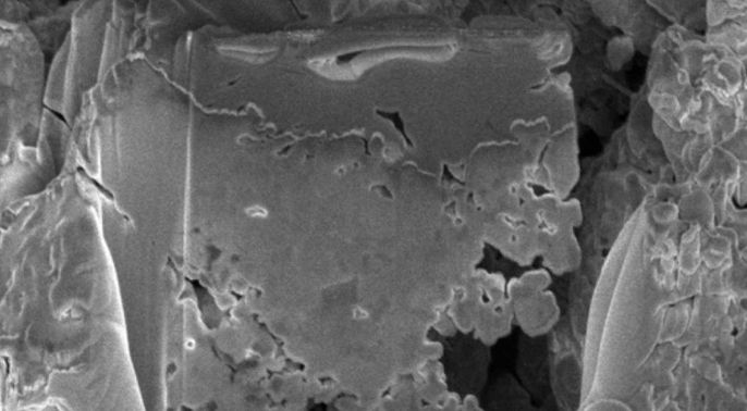
Single slice and view image from 3D dataset
-
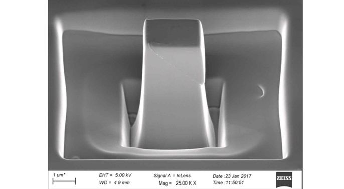
Single crystal Zr micropillar after compression to 5 percent
-
TFS Helios 5 CX help and support
-
Dr Cati Ware
/prod01/channel_3/media/migration/faculty-of-engineering/Cati-Ware-(March-2016)--tojpeg_1499781494737_x4.jpg)
Personal details
Dr Cati Ware Research Facility Assistant (Microscopy)Send email+44 (0) 7872 850079
Location
Department of Materials
Royal School of Mines
Lower Ground Floor, LG05Support with
All Helios NanoLab 600 functions, and SEM and TEM/FIB sample preparation for materials characterisation