The Auriga combines a high resolution SEM to the precision milling and a high resolution FIB.
This SEM features a Schottky field emission gun with Gemini electron column operating between 100 V and 30 kV. This gives spatial resolutions of 1.0 nm. The SEM column is coupled with a Ga+ ion FIB. The FIB column operates between 1 kV and 30 kV with a range of ion beam currents between 1 pA and 20 nA. It also has an imaging resolution of 2.5 nm.
The FIB/SEM is routinely is used for high resolution imaging of biological sample at very low kV to achieve high contrast and 3D reconstruction of distribution of nano-scale microstructures.
High-resolution images are obtained with a standard in-chamber ET detector, and two in-column detectors - a secondary electron detector and an energy selective backscattered electron detector.
A ZEISS gas injection system is fitted, which enables the deposition of platinum to protect the sample surface during ion milling.
Chemical analysis can be carried out on the Oxford Instruments INCA.
Images Auriga
---large--tojpeg_1430819829381_x4.jpg)
Micropillar compression test on two-phase Ti6246, showing slip of and load-displacement-time on the prismatic plane. Test was performed using a nanoindenter (Alemnis)

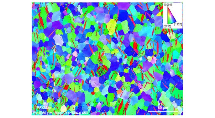
EBSD map of Zircaloy-4 compressed to 8.5% strain along the rolling direction. The IPF colouring shows the orientation of the grains with respect to the plate rolling direction. The red lenticular grains are tensile (T1) twins; the c-axis of the twins are rotated 85 degrees away from the parent grain
Zeiss Auriga Cross Beam help and support
-
Dr Mahmoud Ardakani
/prod01/channel_3/media/migration/faculty-of-engineering/Mahmoud-Ardakani--(Nov-2013)--tojpeg_1499781242792_x4-4.jpg)
Location
Department of Materials
Royal School of Mines
Lower Ground Floor, LG05
